
BL3.2Ua Photoemission Spectroscopy (PES)

Techniques using synchrotron radiation:
-
Soft X-ray and VUV Angle-Resolved Photoemission Spectroscopy (ARPES)
-
Soft X-ray and VUV Angle-Integrated and Angle-Resolved photoemission spectroscopy (SXPS, ARXPS, UPS)
-
Soft X-ray absorption spectroscopy in the total electron yield mode (SXAS, NEXAFS)
Applications:
-
Electronic structural investigation of materials

ARPES End Station

The ARPES end-station consists of photoelectron analysis, sample preparation, and 7 sample slots with linear sample transfer. An angle-resolved electron energy analyzer (VG Scienta, R4000) is installed in the photoelectron analysis chamber. The sample preparation can be carried out in the sample preparation chamber, located above the photoelectron analysis chamber, with the Ar ion sputtering gun (3 keV@ion energy), LEED optics (1 keV), alkali metal evaporator (K, Na, Li), and gas-inlet variable leak valves connected with the roughing pump. A sample can be handled on a 6-axis manipulator, and the temperature-controlled manipulator can be operated down to 8 K. A linear sample-transfer system transfers samples between the vacuum chambers without breaking UHV (⁓1E-10 mbar at low temperature). The MBE chamber consists of a temperature-controlled sample manipulator (RT-1000K@max).
The Vacuum Ultraviolet (VUV) gas-discharged lamp system is available at the ARPES end station. Ultra-pure He (99.999%) gas is excited by high-intensity radio-frequency to generate 21.22 eV photon energy with high intensity (⁓1E12 photons/sec).
XPS End Station

The XPS station performs at room temperature , and the system consists of the hemispherical electron Energy Analyzer (Alpha110) and an Ar ion sputtering gun (3 keV@ion energy). Samples are loaded into the UHV chamber (⁓1E-10 mbar) by using the magnetic linear transfer rod from the first-entry air-lock system, pumped by the turbo-molecular pump, to the XPS analysis chamber, pumped by the 500 l/s ion and titanium sublimation pumps. The residual gas analyzer (100 atomic mass@max) is also available.
BL3.2Ub Photoemission Electron Microscopy (PEEM)

Techniques using synchrotron radiation:
-
Photoemission Electron Microscopy (PEEM), micro/imaging-NEXAFS, XPS, ARPES, Work-Function mapping,
Offline source:
-
LEEM (IV), LEED (IV), and Micro EELS
Applications:
Microscopy for surface, interface, and thin-film research. Electronic structural investigation of surface and interface materials.
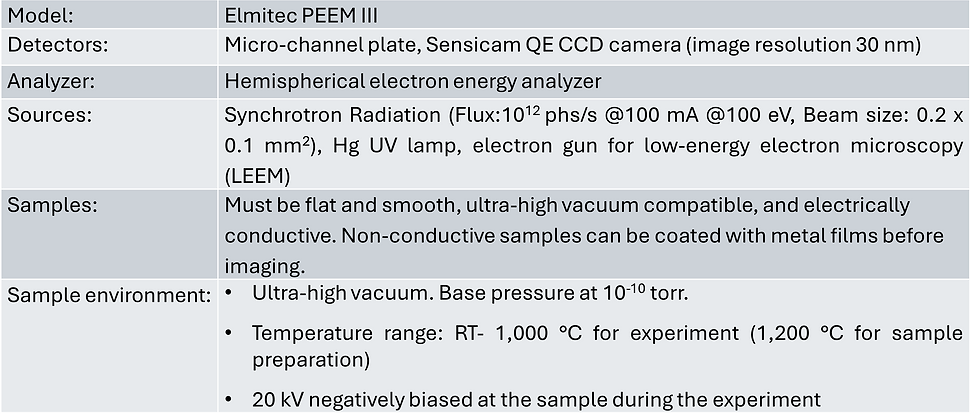
PEEM End Station

current
At PEEM station, a sample is irradiated with monochromatic light from a varied line-spacing plane-grating monochromator. Electrons, created by photoemission and photoabsorption processes, are projected by a set of magnetic lenses onto a micro-channelplate intensifier and a phosphor screen, where the final image forms. By scanning incident photon energy and capturing PEEM image at each energy step, a series of images that contain photoabsorption spectra from nanometer-sized areas of the sample can be obtained. Alternatively, the photon energy is fixed, and an electron energy analyzer is used to determine the photoemission spectrum of photoelectrons emitted from specific areas of the sample.
The electron gun enables structural characterization of crystalline surfaces through Low Energy Electron Microscopy (LEEM), which is highly sensitive to surface topography, atomic step edges, and surface termination. LEEM is typically used to monitor real-time thin-film growth, structural domain formation, gas adsorption, and phase transitions. In reciprocal space mode, the electron gun allows Micro Low-Energy Electron Diffraction (μ-LEED) from regions as small as 0.5 mm using an illumination aperture. By scanning the incident electron energy, LEED intensity curves can be efficiently recorded. It is also possible to select an off-normal LEED spot to form LEEM images in dark-field mode, thereby enhancing contrast between structural islands